
Figure 12.
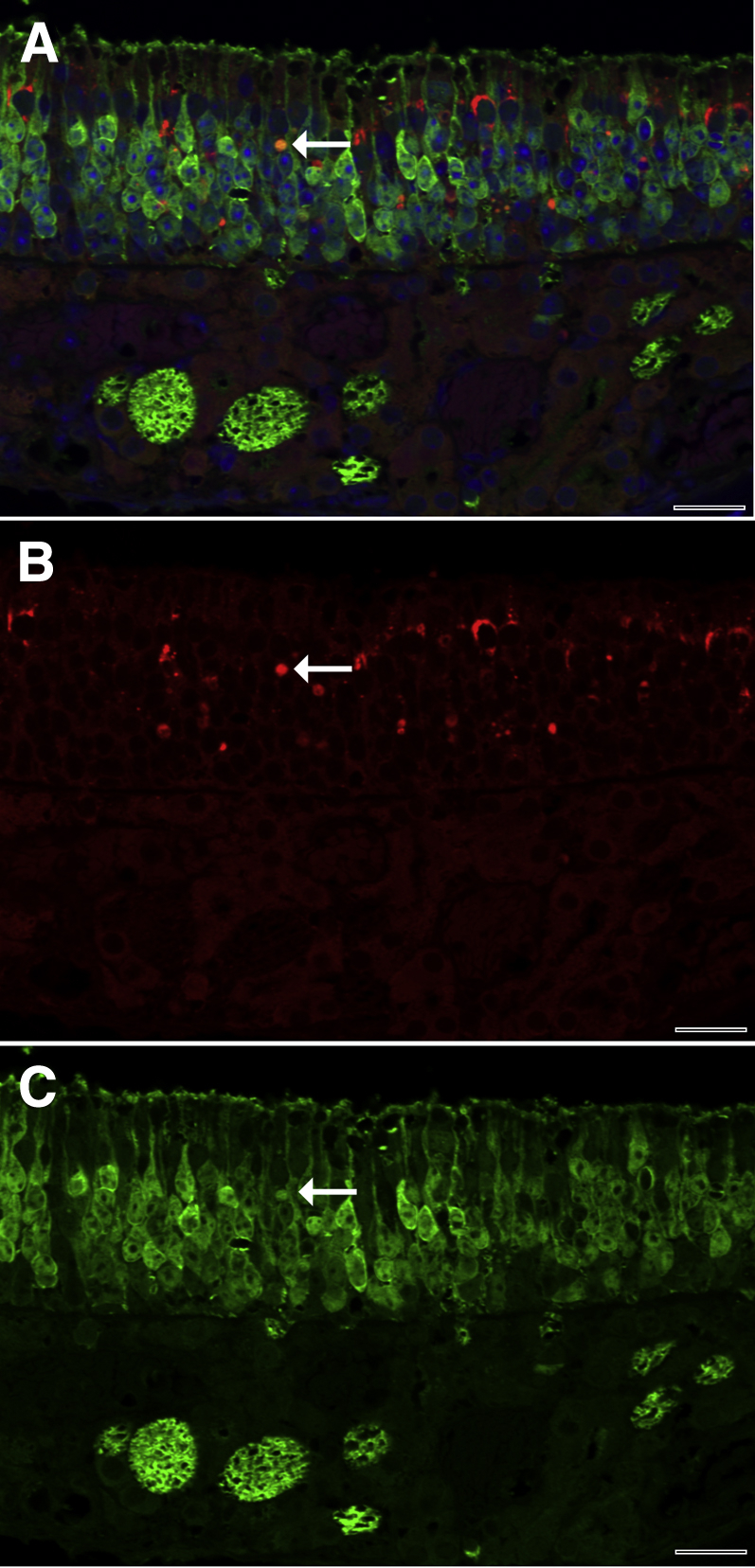
Representative immunofluorescence confocal photomicrograph of SQSTM1 in the neuroepithelium of the mouse nose. A: Merged double-label immunofluorescence demonstrates that SQSTM1 (red) is principally localized between olfactory neurons, which contain OMP (green). Rare cells expressed both SQSTM1 and OMP and were orange (arrow), consistent with neurons containing SQSTM1. B: Red fluorescence demonstrates that foci of SQSTM1 immunoreactivity are abundant and variably sized in the neuroepithelium. The neuron identified in the merged image shows strong SQSTM1 immunoreactivity (arrow). C: Green fluorescence demonstrates immunofluorescence for OMP. The neuron identified in the double-label image shows strong OMP immunoreactivity (arrow). Scale bar = 20 μm (A–C).