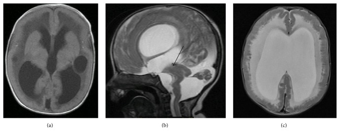
Figure 1.
(a) Axial FLAIR: agyria enlarged ventricle, bilateral periventricle cystic hypodenisty, and delayed myelination of cerebral cortex. (b) Sagittal T2: enlarged lateral and third ventricle. Hypoplastic cerebellum, large posterior fossa CSF fluid space. There is no visible aqueduct (arrow), kinked pontomesencephalic kink. (c) Axial T2-weighted image reveals typical changes related to cobblestone lissencephaly. White matter is diffusely hyperintensity. Enlarged ventricle.

