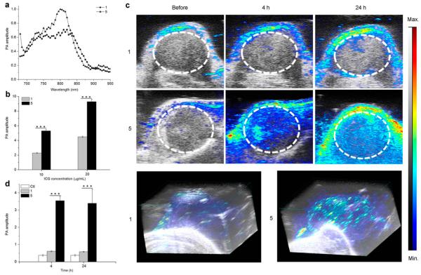
Figure 4.
In vivo in situ enzyme-triggered self-assembly of 5 monitored by photoacoustic imaging. (a) PA spectra of 1 and 5. (b) PA intensity changes of 1 and 5 at different ICG concentrations (1 and 2 in a ratio of 1:100). ***P < 0.001. (c) Representative 2D and 3D PA images of 1 (1, 10 mg/kg, n = 4) and 5 (1, 10 mg/kg; 2, 100 mg/kg, n = 4) on HeLa tumor tissues at 4 or 24 h p.i. The red circles indicate the region of interest in the tumors. (d) PA intensities of tumor tissues at 4 or 24 h p.i. ***P < 0.001.

