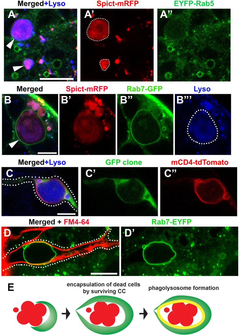
Figure 5. Spict localizes to Rab7-positive phagosomes that encapsulate dying SGs.
(A) Spict-mRFP expressed in CCs (spict-gal4 > UAS-spict-mRFP) does not colocalize with the early endosome marker Rab5. Dying SGs are indicated by arrowheads. Bar: 10 μm. (B) The late endosome marker Rab7 colocalizes with Spict-mRFP and forms a large vesicle encapsulating dying SGs (arrowhead). Dying SG is encircled by dotted line in B”’. Bar: 5 μm. (C) An example of a single CC clone expressing GFP and mCD4-tdTomato (hs-FLP, act > stop > gal4, UAS-GFP, UAS-mCD4-tdTomato) encapsulating dying SGs entirely. A single CC clone is indicated by dotted line. Bar: 10 μm. (D) Rab7-EYFP testis stained for the membrane dye FM4-64 to label the CC plasma membrane, demonstrating that the Rab7-positive vesicle is entirely encapsulated within a single CC. CC boundary is indicated by dotted line. Bar: 10 μm. (E) Model of SG phagocytosis by the surviving CC.

