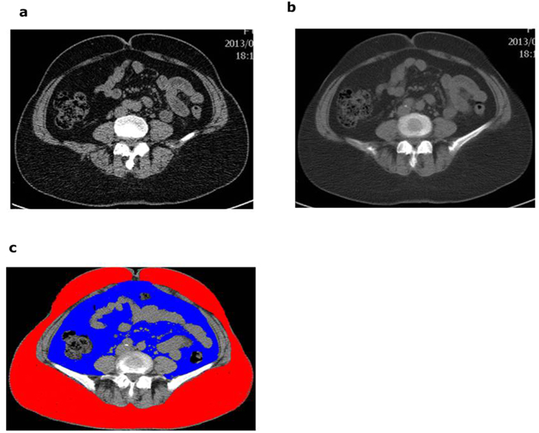
Figure 3. Axial views of an abdominal CT scan.
An image taken through the umbilical plane with slice thickness of 2.5 mm. (b) A smooth image with the slice thickness of 10 mm was reconstructed from four 2.5-mm thick contiguous slices. (c) The SAT and VAT area estimated by AZE Virtual Place software from the reconstructed image ( : SAT,
  : VAT).

