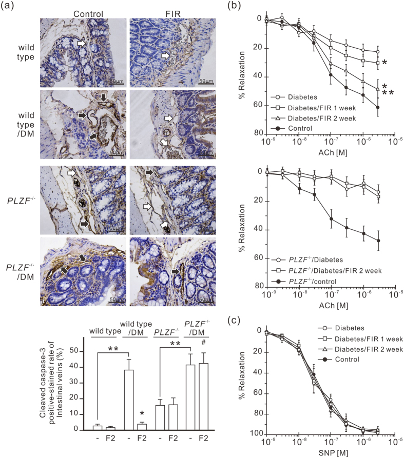
Figure 6. FIR-induced PLZF activation protects vascular endothelium in diabetic mice.
(a) The inhibitory effect of FIR on the expression level of cleaved caspase-3 in the endothelium of intestinal microvessels. Mice were exposed to daily FIR irradiation of 30 min for 2 weeks. The intestines from each mouse were stained by immunohistochemistry with anti-cleaved caspase-3 antibody. The white and black arrows indicate the negative- and positive-stained endothelium of intestinal microvessels respectively. The staining results are also presented as a bar chart form of positive-stained rate of intestinal microvessels. DM, diabetic mice. *P < 0.05 vs. the DM wild-type mice without FIR irradiation. #No significant difference from the PLZF−/−/DM mice without FIR irradiation. **P < 0.05. Scale bar = 50 μm. (b) Acetylcholine (ACh)-induced relaxation in aortic rings of diabetic mice. Diabetic wild-type and PLZF−/− mice were exposed to daily FIR irradiation of 30 min for 2 weeks. (c) Sodium nitroprusside (SNP)-induced relaxation in aortic rings of diabetic mice. ACh- and SNP-induce relaxation of the thoracic aortic rings were measured and expressed as percent of precontraction. Each point of the curve comes from 5 independent experiments with thoracic aortic rings in each group (the mean ± SD). *P < 0.05 vs. DM wild-type mice without FIR irradiation. **P < 0.05 vs. DM wild-type mice with 1-week FIR irradiation.

