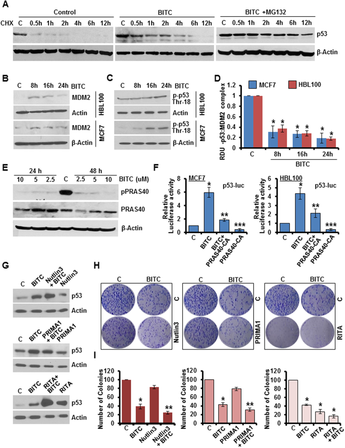
Figure 2. BITC induces stabilization of p53 via PRAS40 modulation. BITC is a potent activator of p53.
(A) MCF7 breast cancer cells were treated with 20 μg/ml cycloheximide (CHX) in the presence of 2.5 μM BITC or 2.5 μM BITC and 20 μM MG132 and lysed at various time points as indicated. Total protein lysates were immunoblotted for p53 expression. Actin was used as control. (B,C) MCF7 and HBL100 cells were treated with 2.5 μM BITC as indicated and total protein lysates were immunoblotted for MDM2 and phospho-p53-Thr18 expression. (D) MCF7 and HBL100 cells were treated with 2.5 μM BITC, whole cell lysates were immunoprecipitated using MDM2 antibodies and purified immunoprecipitates were examined for p53 expression. IgG was used as control. Bar diagram shows quantitation of western blot signals from multiple independent experiments. (E) MCF7 cells were treated with various concentrations of BITC as indicated for 24 and 48 hours, total protein lysates were immunoblotted for phospho-PRAS40 and total PRAS40 expression. β-Actin was used as control. (F) MCF7 and HBL100 were transfected with p53-luc and/or constitutively-active PRAS40 (PRAS40-CA) and treated with 2.5 μM BITC as indicated followed by luciferase assay. (G) MCF7 cells were treated with 2.5 μM BITC, 5 μM Nutlin3, 25 μM PRIMA1, 0.05 μM RITA alone or in combination as indicated, cell lysates were examined for p53 expression. (H,I) MCF7 cells were treated as in G and subjected to clonogenicity. Bar-diagram shows percentage of number of colonies. *P < 0.001, compared with controls; **P < 0.005, compared with Nutlin3 or PRIMA1 alone; denoted with the letter “C”.

