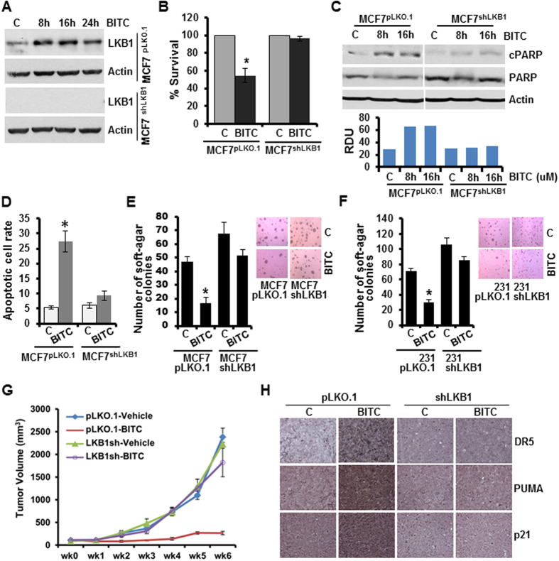
Figure 6. LKB1 is involved in BITC-mediated growth-inhibition and apoptotic-induction in breast cancer cells.
(A) Total protein lysates of LKB1-depleted (LKB1shRNA) and vector control (pLKO.1) MCF7 cells were immunoblotted for the expression of LKB1. MCF7-LKB1shRNA and MCF7- pLKO.1 cells were treated with 2.5 μM BITC and total protein lysates were examined for LKB1 expression in an immunoblot assay. β-actin was used as control. (B) MCF7-LKB1shRNA and MCF7- pLKO.1 cells were treated with 2.5 μM BITC and subjected to XTT assay. *p < 0.01, compared with untreated controls. Vehicle-treated cells are denoted with the letter “C”. (C) Total protein lysates from MCF7-LKB1shRNA and MCF7- pLKO.1 cells treated with 2.5 μM BITC for 8 and 16 hours were immunoblotted for the expression of cleaved PARP and total PARP. β-actin was used as a control. Bar diagram shows quantitation of western blot signals from multiple independent experiments. (D) MCF7-LKB1shRNA and MCF7- pLKO.1 cells were treated with 2.5 μM BITC and subjected to Annexin V/PI staining. *p < 0.01, compared with untreated controls. (E) MCF7-LKB1shRNA and MCF7- pLKO.1 cells were treated with 2.5 μM BITC and subjected to soft-agar colony formation assay. *p < 0.01, compared with untreated controls. (F) MDA-MB-231-LKB1shRNA and MDA-MB-231-pLKO.1 cells were treated with 2.5 μM BITC and subjected to soft-agar colony formation assay. *p < 0.01, compared with untreated controls. (G) LKB1-depleted (LKB1shRNA) and vector control (pLKO.1) MDA-MB-231 cells derived tumors were developed in nude mice and treated with vehicle and BITC. Tumor growth was monitored by measuring the tumor volume for 6 weeks. (n = 8–10); (p < 0.001), pLKO.1+ BITC compared with LKB1shRNA + BITC. (H) Tumors from LKB1shRNA+Vehicle, pLKO.1+Vehicle, LKB1shRNA + BITC, and pLKO.1+ BITC groups were subjected to Immunohistochemical (IHC) analysis using p21, PUMA and DR5 antibodies.

