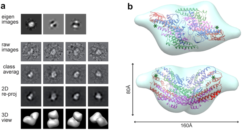
Figure 3. Three dimensional reconstruction of the hGINS double tetramer.
(a) Outline of the 3D reconstruction procedure. Eigen-images indicate the presence of 2-fold symmetry. (b) Double tetramer EM map with a fitted crystallographic dimer12 (PDB ID: 2EHO, chains E to L). The green asterisks indicate the location of the last residue of Psf1.

