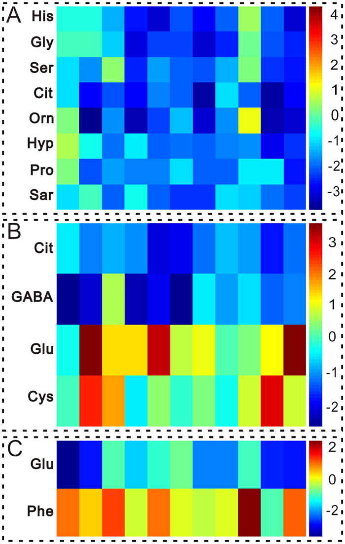
Figure 5. Heat-map for key amino acids change color-scaled with z-scores for the human plasma where the warm (red) colored bars denote elevation of amino acids levels, and cold colored (blue) ones indicate a decrease compared to the CHD patients.

Every amino acid is delegated by a single row of colored boxes, but columns correspond to different samples. Each value was standardized on the basis of the controls (A,B) CHD patients; (C) Chronic AD)) with subtracting mean and dividing by the standard deviation of controls (CHD patients). (A) Acute AD group versus CHD patients; (B) Chronic AD group versus CHD patients. (C) Acute AD group versus Chronic AD group.
