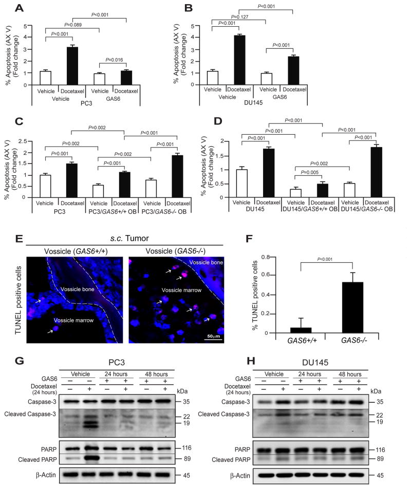
Fig. 4. GAS6 expressed by osteoblasts contributes to the protection of PCa cells from docetaxel-induced apoptosis.
Examination of % apoptosis in (A) PC3 and (B) DU145 cells following the docetaxel treatment with the presence or absence of GAS6 by FACS analysis using Annexin V staining. Examination of % apoptosis in (C) PC3 and (D) DU145 cells in cocultured PC3 cells with GAS6+/+ OB or GAS−/− OB following the treatment of docetaxel. (E) Apoptotic tumor cells (red, white arrows) in the tumor established PC3 cells within GAS6+/+ vossicles or GAS6−/− vossicles as evaluated by TUNEL staining. Blue, DAPI nuclear stain. Bar=50μm. (F) Quantification of % apoptotic cells from Fig. 4E. Docetaxel-induced apoptotic signaling, Caspases-3 and PARP in (G) PC3 cells and (H) DU145 cells following the treatment of docetaxel with the presence or absence of GAS6 were evaluated by Western blots. Data in Fig. 4A–D, F are representative of mean with s.d. (Student’s t-test).

