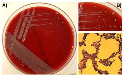
Fig. 2.

(A): Morphology of Staphylococcus sciuri on Columbia blood agar plates. (B): The zoom in indicates white to light grey staphylococcal colonies without any haemolytic activity. (C): Gram-stain visualized Gram-positive, coccoid bacteria, clustered in grape-like structures.
