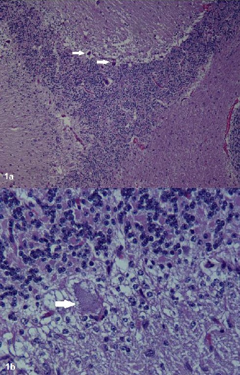
Fig. 1.

Histological examination of cerebellar sections after staining with hematoxylin and eosin. a: Cerebellum of the CA affected foal 10 x: Almost complete absence of Purkinje cells. The remaining Purkinje cells are shrunken and hyperchromatic. b: Cerebellum of the CA affected foal 40 x.
