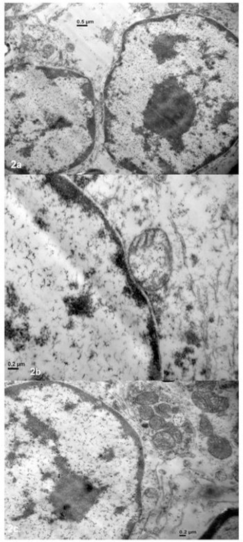
Fig. 2.

Electron microscopy (JEM 1200EX II, Jeol) of a cerebellar section from the CA-affected foal (Camera ES500W Erlangshen C). (a): Morphological characteristics of apoptotic cells (0.5 µm). (b-c): degenerate mitochondria, pale, swollen and vacuolated Purkinje cells (0.2 µm).
