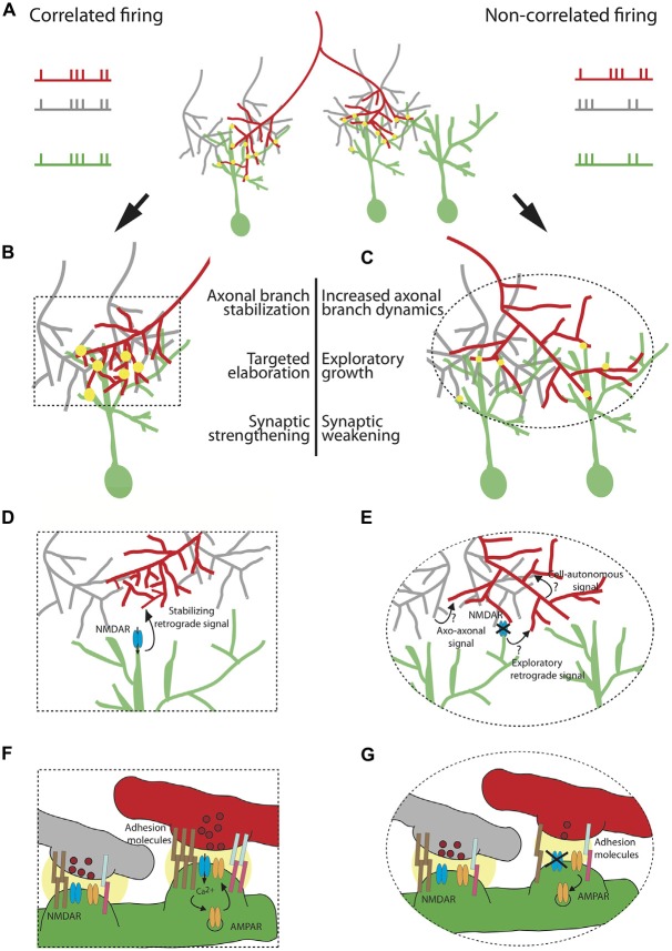
Figure 1.
Cellular and molecular mechanisms underlying the instructive role of patterned neuronal activity in retinotectal map refinement. A retinal ganglion cell (RGC) axon of interest (red) synapses onto tectal neuron dendrites (green) in (A) The red axon (left branch) is co-active with its neighboring RGCs (gray) and its firing pattern is therefore correlated with the firing pattern of the tectal neuron on which it synapses. The right branch of the red axon is not co-active with its neighbors and its firing pattern is not correlated with the firing pattern of its synaptic partner. Synapses are depicted as yellow circles. Synaptic strength is represented by the size of the yellow circle. (B) Correlated firing patterns of an RGC with its partnering tectal neuron instruct an increase in synaptic strength and stabilization of the axonal arbor allowing for local targeted arbor elaboration as new branches tend to form at existing synapses. (C) Non-correlated firing of an RGC with its neighboring axons and its postsynaptic partner instructs synaptic weakening and an increase in axonal branch dynamics, accompanied by exploratory growth in search of a better partner. The effects of patterned neuronal activity on structural remodeling and synaptic efficacy are schematized in (D–G), respectively. (D) Shows a zoom-in of the area of the box in (B) depicting a “stabilizing retrograde signal” downstream of activation of N-methyl D-aspartate receptor (NMDAR) which encodes axonal branch stabilization and targeted elaboration. (E) Zoom-in of the ellipse in (C) Plausible mechanisms instructing axonal exploratory growth and branch destabilization due to the lack of correlated firing with the neighboring RGC inputs and the postsynaptic partner include: 1. Axo-axonal signal, released by the firing neighboring inputs (gray); 2.“Exploratory retrograde signal”, unmasked by the inactivation of NMDAR; 3. Cell-autonomous activity-dependent signal, released by the red neuron or acting intracellularly. (F) Zoom-in depicting molecular mechanisms underlying synaptic strengthening and stabilization of the red axon from (B). Correlated firing of the RGC axon (red) with its neighboring axons (gray) and the tectal neuron (green) results in both release of glutamate containing vesicles (dark red) and postsynaptic depolarization. This satisfies the conditions required for release of the Mg2+ block from the channel pore of the NMDAR (blue), allowing for cation influx. Ca2+ triggers a molecular cascade resulting in insertion of more AMPAR (orange) in the membrane. A retrograde signal downstream of NMDAR encoding higher probability of vesicle fusion in the red axon (depicted as higher number of vesicles in dark red). Increase in synaptic efficacy is accompanied by strong homophilic (brown) and heterophilic interactions of adhesion molecules (light blue and pink). The strength of the interaction is represented by the number of pairs of adhesion molecules. (G) Zoom-in showing the change is synaptic efficacy in (C). Non-correlated firing of the red axon with its neighbors and its partnering tectal neuron prevents opening of the channel pore of NMDAR, leading to AMPAR endocytosis, lower probability of glutamate release and weaker interaction of adhesive molecules.

