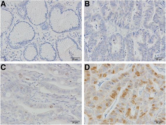
Fig. 2.

Representative photomicrographs of immunostaining for targeting protein for Xenopus kinesin-like protein 2 (TPX2), demonstrating a negative staining in normal gastric epithelium, b negative staining in primary gastric carcinoma, c positive staining graded as low (<5% of tumor cells stained), and d positive staining graded as high (≥5% of tumor cells stained). TPX2 protein was mainly localized within the nuclei of tumor cells. Images were captured at ×200 magnification
