
Fig. 4.
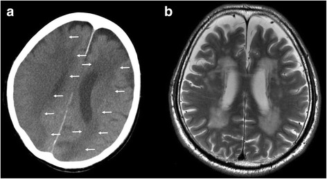
The postmortem CT and antemortem MRI (Case 2). a The postmortem CT image of the brain 2 h after death. LDAs were found widely, resulting in a diagnosis of cerebral infarction (arrows). (with permission [6]) b Antemortem MRI, T2-weighted image showing no cerebral infarction. The brain MRI was taken 1 year 5 months before death