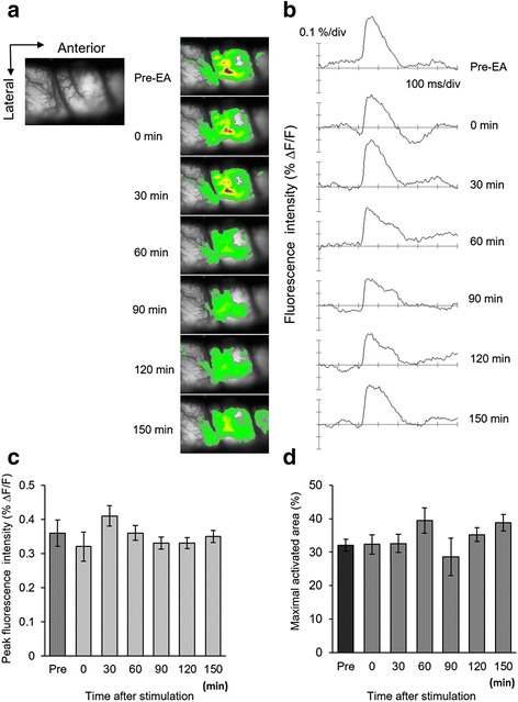
Fig. 6.

Spatiotemporal activities of sham EA stimulation in neuropathic rats. Sham EA stimulation was characterized by EA stimulation at nonacupoint locations (a) Each activated signal in pre- or post-sham EA stimulation was detected after electrical stimulation of the hind paw in nerve-injured rats. After sham EA stimulation, the enlarged and propagated activated area did not reduce. b The peak activated fluorescence signals did not change after sham EA stimulation. c The average of peak fluorescence has shown continuance activated pattern. d Time-dependent observed maximal activated area in S1 cortex is presented (n = 5)
