Figure 4.
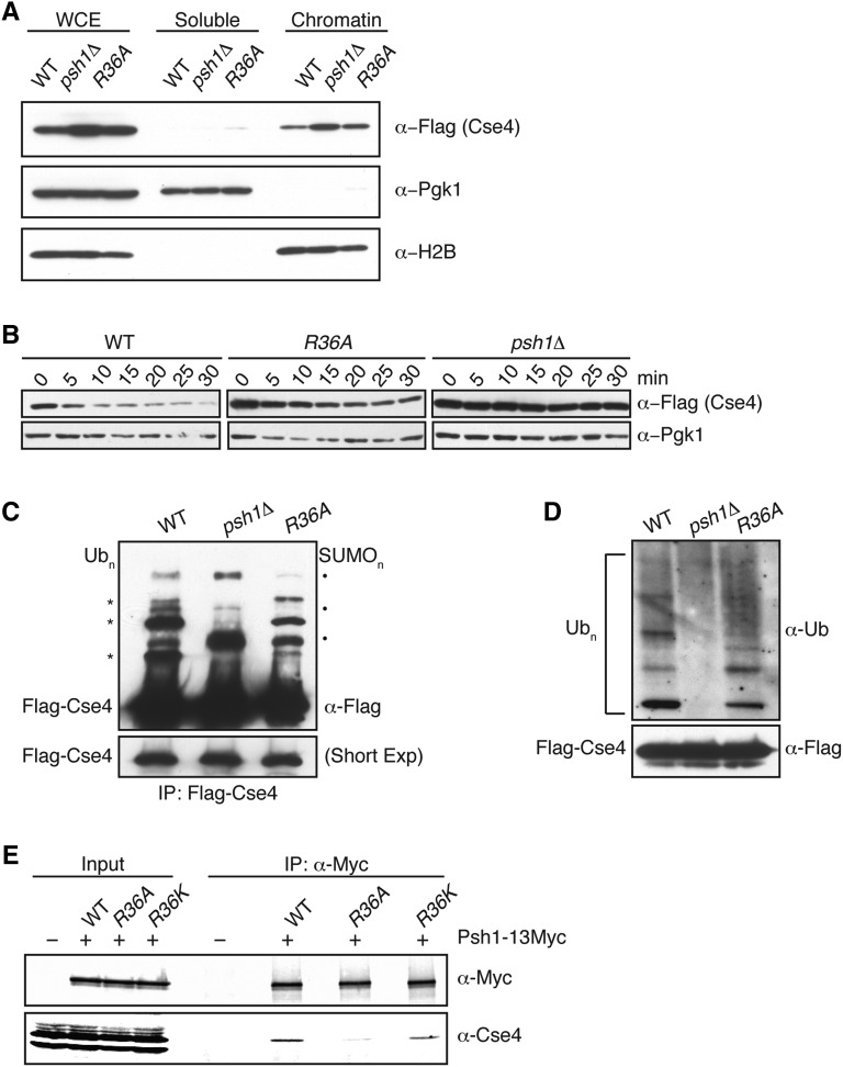
H4-R36A cells have defects in CENP-ACse4 degradation. (A) WCE from WT (SBY10025), psh1∆ (SBY10590), and H4-R36A (SBY9397) cells expressing pGAL-3Flag-CSE4 were fractionated into soluble and chromatin fractions. 3Flag-Cse4 levels were monitored in each fraction with α-Flag antibodies. Pgk1 and H2B are markers of the soluble and chromatin fractions, respectively. Note that a longer exposure of the blot for CENP-ACse4 is included in Figure S3, which shows CENP-ACse4 in the soluble fraction. (B) WT (SBY10025), H4-R36A (SBY9397), and psh1∆ (SBY10590) cells expressing pGAL-3Flag-CSE4 were grown in galactose, protein synthesis was inhibited by cycloheximide addition at time zero, and lysates were monitored for 3Flag-Cse4 levels at the indicated time points with α-Flag antibodies. Pgk1 served as a loading control. (C) WT (SBY10025), psh1∆ (SBY10590), and H4-R36A (SBY9397) cells expressing pGAL-3Flag-CSE4 were grown in galactose and 3Flag-Cse4 was immunoprecipitated with α-Flag antibodies. Unmodified 3Flag-Cse4 and its higher-mobility species (Ubn) were detected on immunoblots by probing with α-Flag antibodies, these bands are marked on the left by asterisks. Note that SUMO conjugates appear on Cse4 in the absence of ubiquitylation (SUMOn), these bands are marked on the right by solid dots (Ohkuni et al. 2016). The lower panel shows a shorter exposure of the immunoblot to display the levels of unmodified 3Flag-Cse4 in the immunoprecipitates and serves as a loading control. (D) An immunoblot probed with α-ubiquitin antibodies of the α-Flag immunoprecipitates isolated from the strains denoted in (C) reveals the ubiquitin conjugates of 3Flag-Cse4. The lower panel shows the levels of unmodified 3Flag-Cse4 by immunoblotting with α-Flag antibodies. (E) Psh1-13Myc was immunoprecipitated from WT (SBY15335), H4-R36A (SBY15624), and H4-R36K (SBY16468) cells, and immunoblots were probed with α-Myc and α-Cse4 antibodies. Cells expressing untagged Psh1 (SBY3) served as a control. The Cse4/Psh1 ratio ± 1 SEM was calculated vs. WT for H4-R36A (0.5 ± 0.03) and H4-R36K (3.0 ± 0.6) from three biological replicates. IP, immunoprecipitation; WCE, whole cell extract; WT, wild-type.

