Figure 5.
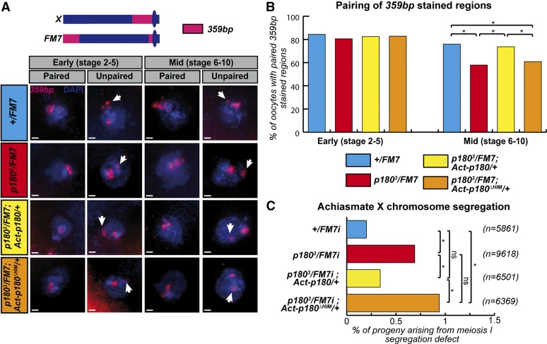
The HIM domain participates in heterochromatin-mediated pairing in germ cells. (A) FISH staining on oocytes allows the detection of X pericentric regions (359 bp) and total DNA (DAPI) in females of the indicated genotype. Based on previously described detection of synaptonemal complex components (Resnick et al. 2009), two different regions of the ovariole were analyzed: the early region from vitelline stage 2–5 in which the synaptonemal complex, maintaining aligned homologs, is fully assembled; and the middle region containing egg chambers from stage 6–10, in which the synaptonemal complex is undergoing disassembly and therefore likely requires additional mechanisms to maintain pericentric interactions. Scale bar represents 1 μm. (B) Pairing of 359-bp regions of homologous X chromosomes was quantified in females of the indicated genotype. Numbers of oocytes scored for stages 2–5 (early): +/FM7i, 64; p1803/FM7i, 68; p1803/FM7;Act-p180/+, 80; and p1803/FM7i;Actp180ΔHIM/+, 126. Number of oocytes scored for stages 6–10 (late): +/FM7i, 75; p1803/FM7i, 63; p1803/FM7;Act-p180/+, 57; and p1803/FM7i;Actp180ΔHIM/+, 99. (C) Rate of exceptional oocytes arising from X chromosome-segregation defects during meiosis I, measured in females of the indicated genotype. In this experiment, due to heterozygosity of the females for the FM7i balancer chromosome, X chromosomes fail to recombine and therefore segregate according to the homologous achiasmate system. Differences are indicated to be statistically significant (*) or not significant (ns) using Fisher’s exact test (P ≤ 0.05).

