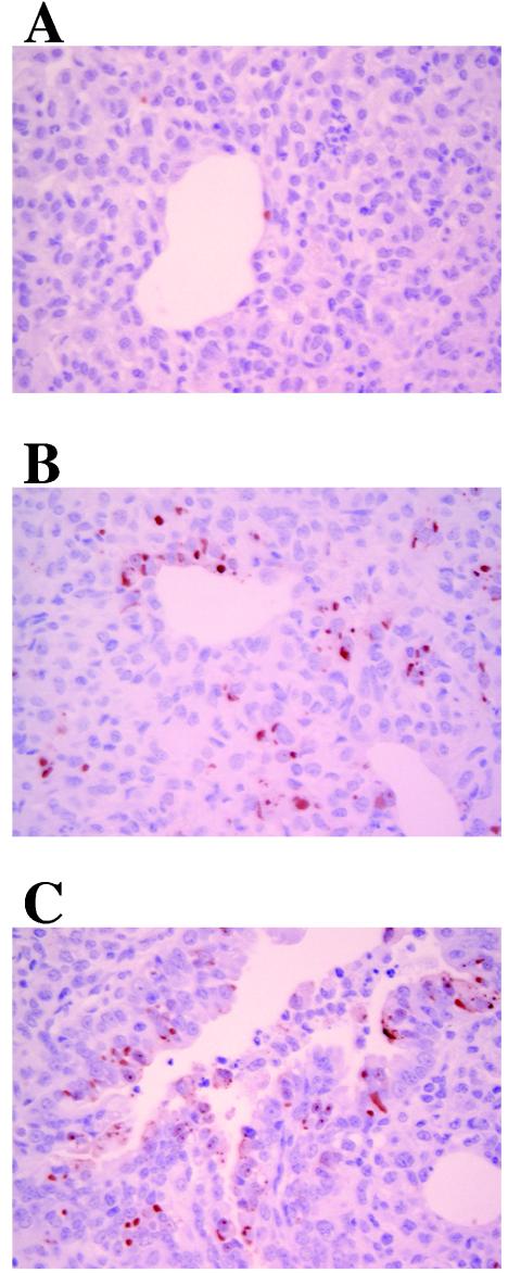
FIG. 6.
Immunohistochemistry for PIV3 in the PIV3 group (A), PIV3/Ad group (B), and PIV3/Ad/HBD6 group (C). The PIV3/Ad and animals in the PIV3/Ad/HBD6 group had increased cellular staining compared with that for the animals in the PIV3 group. No staining was detected for the animals in the control group (not shown). Magnifications, ×400.

