Figure 6.
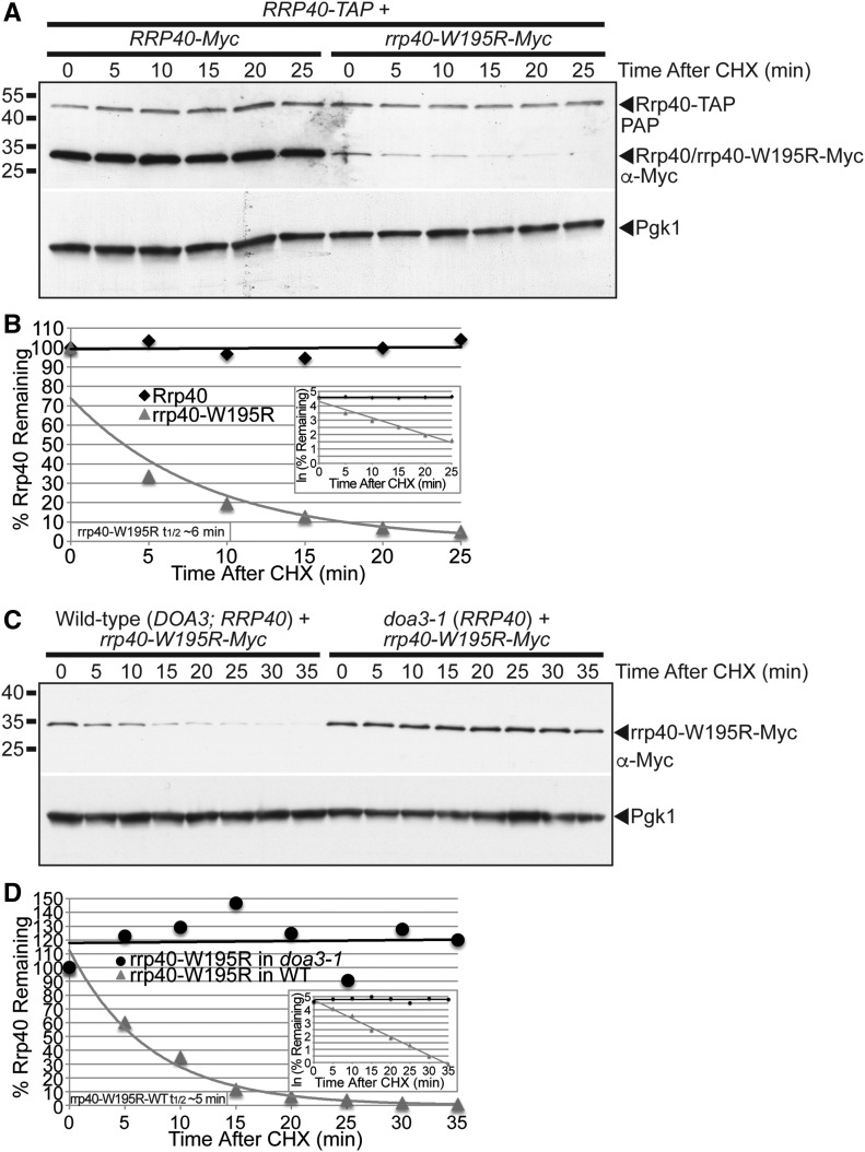
The rrp40-W195R variant is unstable and degraded by the proteasome in S. cerevisiae cells coexpressing wild-type Rrp40. (A) The level of rrp40-W195R-Myc variant decreases more rapidly than wild-type Rrp40-Myc over time in cells coexpressing wild-type Rrp40-TAP at 30°. RRP40-TAP cells expressing wild-type Rrp40-Myc or rrp40-W195R variant at 30° were treated with CHX to inhibit translation. Samples were collected over time (0–25 min) and analyzed by immunoblotting with anti-Myc antibody to detect Myc-tagged proteins (Rrp40/rrp40-W195R-Myc), peroxidase anti-peroxidase (PAP) antibody to detect Rrp40-TAP, and anti-Pgk1 antibody to detect Pgk1 as a loading control. (B) The immunoblot shown in (A) was quantitated to plot the percentage of Rrp40-Myc and rrp40-W195R-Myc protein remaining at each time point relative to inhibition of translation at time 0 in RRP40-TAP cells. The inset graph shows the natural logarithm (ln)-transformed percentages of Rrp40 and rrp40-W195R at time points 0–25 min fitted with linear least-squares fit lines to determine the decay rate constant (k) for each protein. The inset half-life (t1/2) of rrp40-W195R in RRP40-TAP cells at 30° of ∼6 min was calculated from the decay rate constant using the equation t1/2 = ln(2)/k (Belle et al. 2006). (C) The level of rrp40-W195R-Myc variant is increased in doa3-1 proteasome mutant cells coexpressing wild-type Rrp40 at 37°. Wild-type or doa3-1 cells expressing rrp40-W195R-Myc protein at 37° were treated with CHX. Samples were collected over time (0–35 min) and analyzed by immunoblotting with anti-Myc antibody to detect rrp40-W195R-Myc protein, and anti-Pgk1 antibody to detect Pgk1 as a loading control. (D) The immunoblot shown in (C) was quantitated to plot the percentage of rrp40-W195R-Myc protein at each time point in wild-type and doa3-1 mutant cells. The inset graph shows the natural logarithm (ln)-transformed percentages of rrp40-W195R at time points 0–35 min in wild-type or doa3-1 cells fitted with linear least-squares fit lines to determine the decay rate constant (k) for each protein. The inset half-life (t1/2) of rrp40-W195R in wild-type cells at 30° of ∼5 min was calculated as described in (B). Further details on the measurement of the protein band intensities and calculation of the protein half-lives are described in Materials and Methods. Quantitation is for the specific experiment shown, but is representative of multiple experiments.

