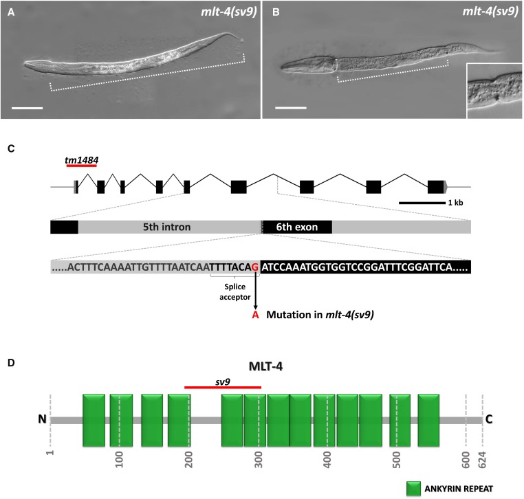
Figure 1.
Molting defects in mlt-4(sv9) mutant animals. (A, B) DIC images of sv9 homozygous larvae entrapped within two layers of cuticle. Dotted line indicates the extent of the constricted region containing both the old and new cuticle layers. A larva in which only the head region is free of old cuticle is shown in (A), whereas the larva in (B) displays a classic corset phenotype in which both the head and tail regions have released the old cuticle. The inset in (B) shows an enlargement of the posterior constriction. (C) Schematic representation of the mlt-4 gene; the affected region with the splice acceptor site that is mutated in the sv9 allele is enlarged. The red line marks the location of the tm1484 deletion. (D) Schematic illustration of MLT-4 with annotated predicted ankyrin repeats (green boxes). The red line indicates the region that would be affected by mis-splicing in sv9 mutants. Numbers specify positions of amino acids. Bar, 50 µm (A, B).

