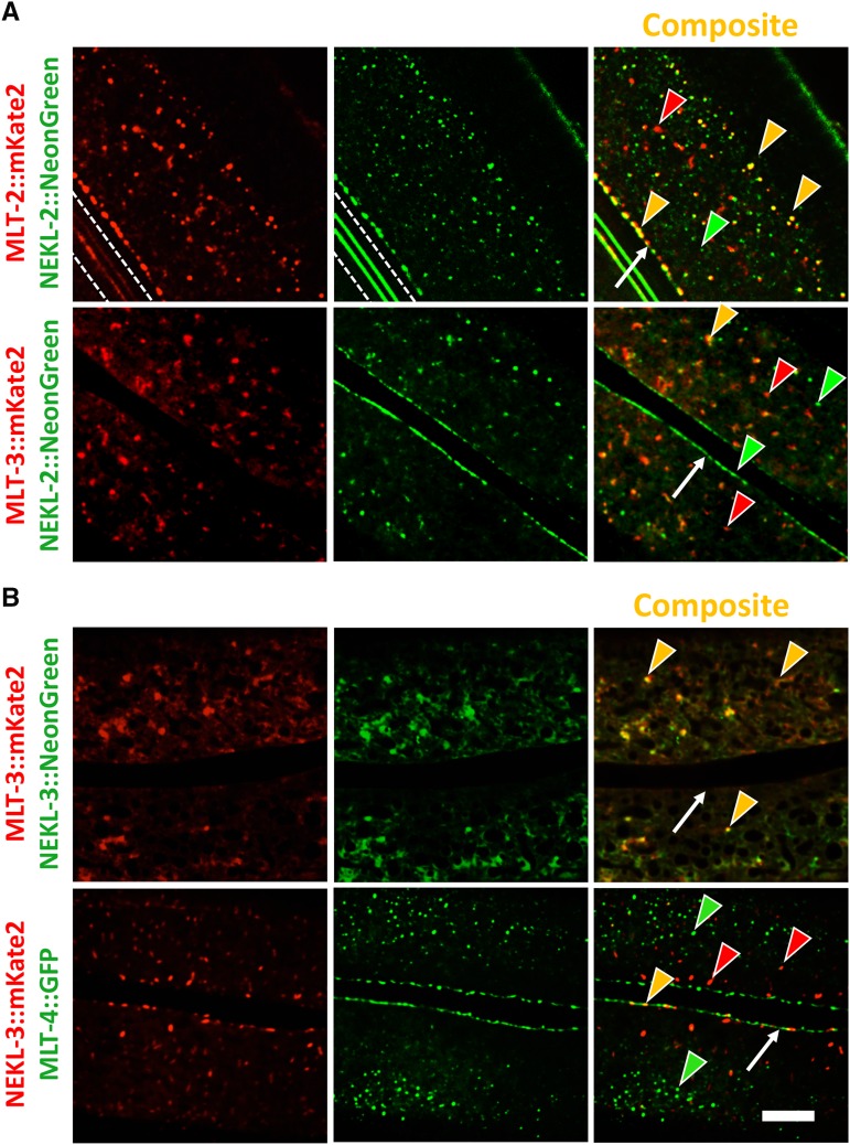
Figure 7.
Colocalization of NEKL and MLT proteins. (A) Colocalization analyses of MLT-2::mKate2 and MLT-3::mKate2 with NEKL-2::NeonGreen showing extensive overlap of MLT-2 and NEKL-2 puncta throughout the hyp7 apical surface including the region adjacent to the seam cell. Though less frequent, some NEKL-2 puncta colocalized with MLT-3. White dashed lines in the first two panels demarcate autofluorescent alae. (B) NEKL-3::NeonGreen apical accumulations extensively overlapped with MLT-3::mKate2. In contrast, NEKL-3::mKate2 did not colocalize with MLT-4::GFP puncta. Red and green arrowheads indicate representative puncta that express only one marker, whereas yellow arrowheads indicate colocalization. White arrows indicate hyp7–seam cell boundaries. Bar, 5 µm.

