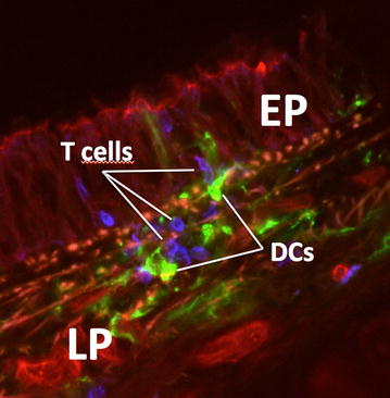
Fig. 1.

Interactions of DCs and T cells in the airway mucosa visualized by laser scanning microscopy. Micrograph shows a scanned area from a cross section through a trachea (rat). T cells (CD3, blue) are visible closely interacting with DCs (MHC class II, green) inside the airway epithelium (EP) and the lamina propria (LP)
