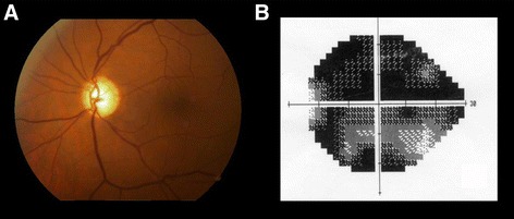
Fig. 3.

Disc cupping and pallor (a) associated with compressive lesion of the intracranial portion of the left optic nerve caused by a dolichoectatic internal carotid artery. Reduced visual acuity, loss of the central visual field (b) and neuroretinal rim pallor indicated the need for neuroimaging investigation
