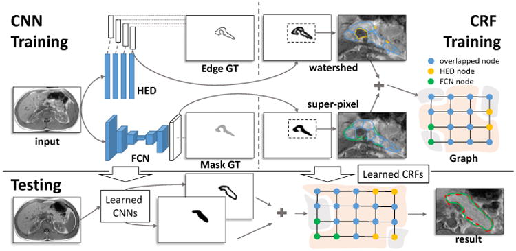
Fig.1.

The framework of our approach. CNN Training: CNN models are trained for pancreatic tissue allocation (the FCN model) and boundary detection (the HED model); CRF Training: A CRF model is learned based on the candidate regions that detected by CNN models. Testing: The segmentation begins with CNN models, and then will be further refined by the CRF model. The result of testing and the corresponding human annotation are displayed with the green and red dashed curves, respectively.
