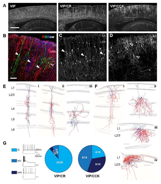
Figure 3. Intersectional targeting of VIP subpopulations.
(A–C) Representative cortical sagittal sections from VIP-Flp;tdTomato-FRT (VIP), VIP-Flp;CR-Cre;Ai65 (VIP/CR) and VIP-Flp;CCK-Cre;Ai65 (VIP/CCK) mice. (A) Lower magnification view of reporter expression in sagittal sections. (B) Triple color labeling by RGBow reporter via VIP-Cre activation revealed several morphologically distinct neuron types intermingled in superficial layers. (C–D) Higher magnification images of boxed area in (A). Arrow heads: neurons with vertically restricted axons and dendrites. Open arrow heads: neurons with multipolar, small basket cell like morphology. (E–F) Neurolucida reconstruction of biocytin filled neurons recorded in VIP/CR neocortex (E) and VIP/CCK neocortex (F). Axons were traced in red and dendrites in blue. (G) Current clamp recordings in somatosensory cortex of VIP/CR and VIP/CCK mice show different proportions of cells with various intrinsic firing patterns. Representative recording traces are shown on the left. Irregular spiking cells (IS) are characterized by action potentials elicited with irregular intervals at near threshold. Fast adapting cells (fAD) are characterized by their failure to fire throughout the 600 ms step during suprathreshold depolarization. Burst non-adapting cells (bNA) exhibit bursts of two or three spikes, followed by regular spiking. Proportions of each firing pattern in the two intersection lines are shown as pie charts on the right. Scale bar in A: 500μm. Scale bar in B–F: 100μm. (See also Figure S4, Table S2–5.).

