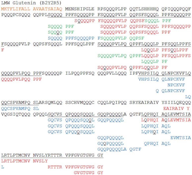
Figure 6.
Sequence coverage of an exemplar LMW glutenin subunit (B2Y2R5). Peptide sequences unique to the QTOF with ion mobility incorporated DIA are shown in red, sequences unique to the LTQ with DDA in blue with shared peptides shown in green. Modified amino acids are highlighted in black; with black Q's representing deamidated glutamine. When the residue is black and underlined this represents that the modification could occur at multiple points within the sequence, and its exact location is unknown. The signal peptide is highlighted in orange. The underlined sequence represents the regions of the sequences covered by the peptides from either the QTOF or LTQ data.

