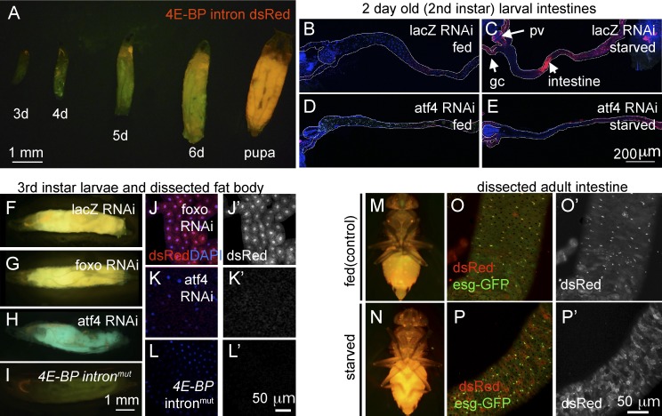
Figure 4.
4E-BP intron dsRed reporter marks cells with active ATF4 signaling. Shown are 4E-BP intron dsRed reporters in larvae (A–L) and adult flies (M–P). Unless specified otherwise, 4E-BP intron dsRed is marked in red and GFP in green. (A) The reporter expression during larval development (numbers indicate days after egg laying). (B–E) Second-instar larval intestines. Anterior is to the left, and posterior is to right. The outlines of the intestines are marked in white. Before dissection, the larvae were either well fed (B and D) or deprived of amino acids for 6 h (C and E). These intestines were expressing RNAi lines against control lacZ (B and C) or atf4 (D and E). dsRed reporter induction in response to amino acid deprivation (C) can be seen in the proventriculus (pv), gastric caeca (gc), and the intestine (arrows), which is suppressed when atf4 is knocked down (E). (F–I) Third-instar larvae show inherent activation of ATF4 as shown by 4E-BP intron reporter expression. The indicated RNAi lines were driven together with uas-GFP using the fat body–specific cg-Gal4 driver. (J–L) Dissected larval fat bodies containing the 4E-BP intron dsRed reporter. RNAi-mediated knockdown of foxo (J and J′) had no effect, whereas atf4 knockdown blocked the reporter expression (K and K′). (L and L′) The expression of the 4E-BP intronmut reporter, with the putative ATF4-binding sites mutated. (J′, K′, and L′) dsRed-only channels of J, K, and L. Adult flies reared under well-fed conditions (M and O) or those starved overnight (N and P). (M and N) Low-resolution images under dissecting microscopes. (O and P) Dissected intestinal epithelium under confocal microscopy. The posterior intestine, where esg-GFP–positive ISCs are found, was focused. (O′ and P′) dsRed-only channels of O and P. Note that in the epithelium of well-fed flies, the most prominent dsRed signals colocalize with the small nuclear cells that are positive for esg-GFP. In contrast, starved epithelium shows reporter activity spreading to esg-GFP–negative cells with large nuclei.

