Figure 1.
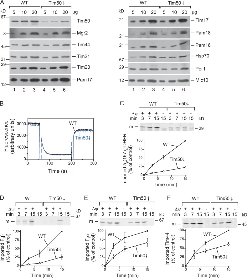
Protein import is impaired in Tim50-depleted mitochondria. (A) Steady-state Western blot analysis of WT and Tim50-depleted mitochondria. (B) Δψ of isolated mitochondria was assessed using the Δψ-sensitive dye DiSC3(5). Fluorescence was recorded before and after addition of valinomycin. (C–F) 35S-labeled precursors were imported into isolated mitochondria, and import stopped at the indicated time points with antimycin A, valinomycin, and oligomycin (AVO). Samples were PK treated and analyzed by SDS-PAGE and autoradiography. Results are presented as mean ± SEM. n = 3. The longest import time of the WT sample was set to 100%. m, mature protein.

