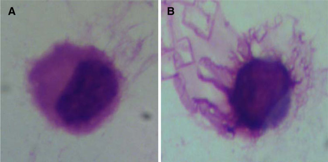
Fig. 1.
Light microscopy of DCs. a Generation of inactivated DCs; MNCs were separated using a Ficoll-Hypaque density gradient; then, MNCs were differentiated into DCs by suspending them in liquid culture medium containing EMEM, 10 % FCS, penicillin (100 U/ml), streptomycin (10 mg/ml), amphotericin B and gentamycin and adding the growth factors rhIL-4 (20 ng/ml) and rhGM-CSF (100 U/ml) to the suspension pulsed with HepG2 cell line lysate in sterile tissue culture tubes that were incubated at 37 °C in 5 % CO2 for 7 days. The medium was changed every 2–3 days. b Generation of activated DCs was done on day 6 by adding 10 ng/ml TNF α; the cultured MNCs were evaluated for morphological changes using cytospin preparation stained with Giemsa. Cells having a large size, copious gray cytoplasm and long cytoplasmic processes were identified as mature DCs

