. 2016 Dec 1;112(1):31–43. doi: 10.1590/0074-02760160270

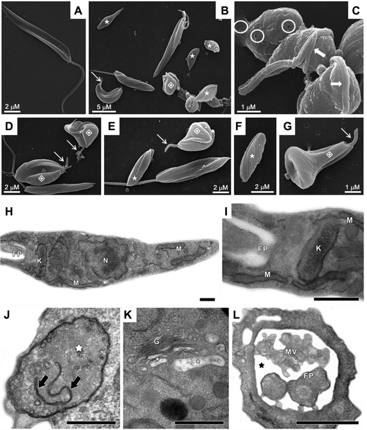
Fig. 1. : scanning electron microscopy (SEM, A-G) and transmission electron microscopy (TEM, H-L) of Phytomonas serpens after treatment with MDL28170. In untreated, control promastigotes (A), note the elongated body and emerging flagellum in SEM. Treatment with the IC50 value of MDL28170 for 48 h (B-G) caused rounding of the parasite body (B, C), shortening (thin arrows) or complete loss (stars) of the flagellum, cell surface discontinuity (circles) and appearance of cells with bizarre shape (diamonds) and with flagellum contorting the cell body (thick arrows). Control parasites in TEM (H, I) showed typical morphology of nucleus (N), mitochondrion (M), kinetoplast (K) and flagellar pocket (FP). The treatment of promastigotes with the IC50 value of MDL28170 for 48 h (J-L) led to the mitochondrial swelling (white star) with membranes in the organelle matrix (black arrows), the disruption of trans-Golgi (G) with peripheral dilation of cisternae (white asterisks), as well as the dilation of flagellar pocket (black star) presenting microvesicules (MV) inside. Bars in TEM: 0.5 µM.

Fig. 1