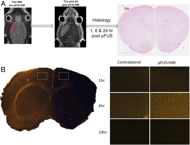
Fig. 1.
BBBD by pFUS+MB. (A) Diagram of the experimental design for pFUS-treated rat brain for histological analysis. T2w MRI was used to target FUS with points (red circles) ∼2 mm in diameter placed in the left frontal cortex anterior to the lateral ventricle. Following pFUS+MB, Gd-enhanced T1w images were obtained. The dashed white line outlines the contrast enhancement. Animals were killed 1, 6, or 24 h after sonication, and brains were collected for histology. No microhemorrhages or macroscopic alterations to morphology were observed on H&E staining. (B) Albumin extravasated through the open BBB with peak parenchymal accumulation occurring at 6 h post pFUS+MB.

