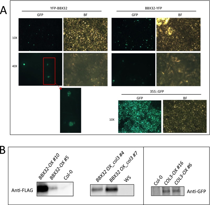
Fig. S4.
Subcellular localization of BBX32 and BBX32 and COL3 protein expression. (A) BBX32 is localized to nuclear speckles. Enlarged image of the nuclei shows the size and number of speckles. 35S::GFP was used as a control. BF, bright field. (B) Protein expression analysis of Flag-tagged BBX32-OX; Flag-tagged BBX32 OX_col3, and GFP-tagged COL3-OX lines. Ten-day-old seedlings grown in 12L:12D conditions were harvested at ZT1, and Western blot analysis was performed. Anti-FLAG and anti-GFP antibodies were used for BBX32-OX; BBX32 OX_col3, and COL3-OX, respectively.

