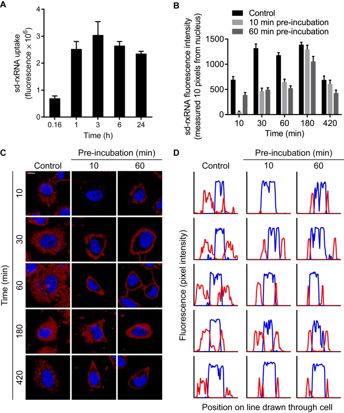
Figure 2.
sd-rxRNA is internalized through a saturable pathway. (A) HeLa cells were treated with 0.5 μM sd-rxRNA-Cy3 and imaged by live confocal microscopy. Uptake was quantified by measuring total fluorescence intensity at specific time points. (B) HeLa cells were either untreated (control) or pre-incubated with 2 μM unlabeled sd-rxRNA for 10 or 60 min. Cells were then treated with 0.5 μM sd-rxRNA-Cy3 and imaged by live cell confocal microscopy. sd-rxRNA fluorescence intensity was measured in a region only 10 pixels from the nucleus. (C) Representative images used for quantification in panels B and D. Blue represents DAPI stain and red represents Cy3-labeled sd-rxRNA. Scale bar = 10 μm. (D) Corresponding plot profiles of lines drawn through images in panel C. Blue lines represent DAPI stain intensity and red lines represent Cy3-labeled sd-rxRNA intensity.

