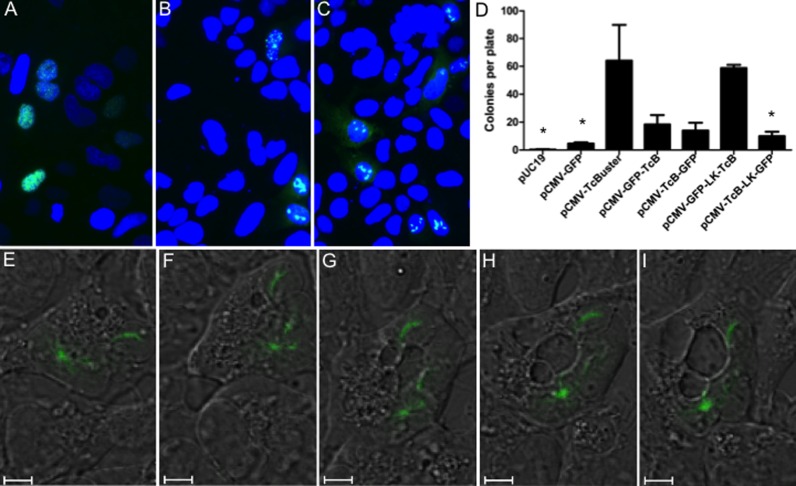
Figure 4.
Live cell imaging of rodlet formation. HEK-293 cells plated onto coverslips in 6-well dishes were transfected with either 1 μg of pCMV-TcBuster-GFP (A), 500 ng of pCMV-TcBuster-GFP + 500 ng of pCMV-TcBuster (B) or 500 ng of pCMV-TcBuster-GFP + 500 ng of pCMV-HA-TcBuster (C). Cells were mounted directly with media containing DAPI to stain the nuclei (blue) to observe GFP expression (green). (D) To test the activity of the GFP-tagged TcBuster transposase (TcB) constructs, with and without a linker (LK), HEK-293 cells were transfected with the transposase plasmid indicated and the pTcBNeo neomycin-resistance transposon plasmid at a 1:9 ratio, n = 3 per group. After 48 h, the cells were split into selection media and colonies were counted after two weeks. Asterisk (*) indicates this group was significantly different than pCMV-TcBuster by Tukey post-test analysis. (E–I) Rodlets were imaged in HEK-293 cells by confocal live cell imaging of DIC (black and white) and GFP (green) 32 h after transfection with 500 ng each of pCMV-TcBuster and pCMV-GFP-LK-TcBuster. A cell is shown at 32:00 (E), 32:30 (F), 33:00 (G), 33:30 (H) and 34:00 (I).

