Figure 5.
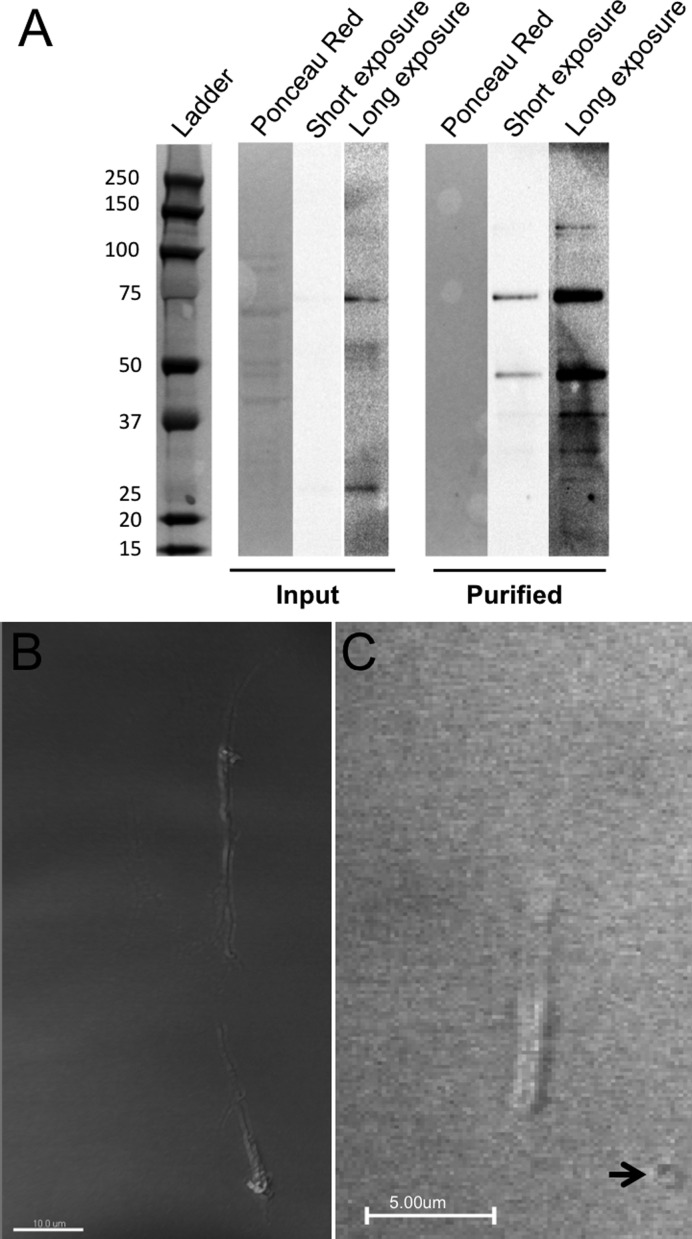
Formation of rodlets from purified transposase ex vivo. Protein was purified with a Flag affinity column from HEK-293 cells transfected via FuGene 6 with pCMV-Flag-TcBuster 48 h prior. (A) Input (unpurified protein lysate) and purified protein were run together, transferred and Flag was detected by chemiluminescent western blot followed by staining with Ponceau Red to determine overall protein amount in each lane. Both a short, unmodified exposure and long exposure optimized for sensitivity are shown. Expected running size for Flag-TcBuster is 75.8 kDa. The size of each band in the ladder is given in kDa. (B and C) Purified Flag-TcBuster formed rodlets in cell-free conditions both in the absence (B) and presence (C) of transposon DNA in a cell-free buffer system. Slides were DIC imaged on a confocal microscope. Larger circular structures (arrow) were present only when DNA was added.

