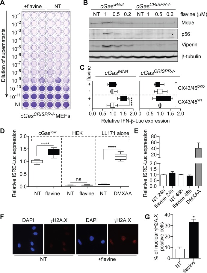
Figure 4.
cGAS-dependent antiviral activity of flavine. (A) Viral titers of cGas-deficient MEFs (19) treated for 72 h with 1 μM flavine and infected for 24 h in biological triplicate with SFV (MOI 2). Viral titers were assayed as in Figure 1. Data shown are representative of three independent experiments in three different clones of cGas-deficient MEFs (19). (B) Dose-response effect of 72 h flavine treatment on MEFs (matched wild-type or cGas-deficient) on the levels of Viperin, Mda5 and p56 by western blot. Data shown are representative of a minimum of two independent experiments. (C) HEK-Sting CX43/45WT and Sting CX43/45DKO cells expressing an IFN-β-Luciferase reporter were co-cultured with MEFs (matched wild-type or cGas-deficient) pre-treated or not with flavine for 24 h. IFN-β-Luciferase expression was reported to the NT condition for each cell line (data presented are averaged from a minimum of two independent experiments in biological triplicate ± s.e.m. and unpaired Mann–Whitney U test is shown). (D) Murine LL171 cells (L929 cells expressing an ISRE-Luciferase reporter) were cultured for 18 h, in the absence (‘LL171 alone’) or presence of HEK cells (wild-type or cGaslow expressing) pre-treated with 1 μM flavine for 24 h. ISRE-Luciferase expression is shown relative to the NT condition for the cGaslow expressing co-culture (data presented are averaged from three independent experiments in biological triplicate ± s.e.m. and unpaired Mann–Whitney U tests are shown). (E) LL171 reporter cells were treated with 1 μM flavine for 24 or 48 h before lysis. NT: not-treated. ISRE-Luciferase activity is shown relative to the NT condition. Data shown are averaged from two independent experiments in biological triplicate (± s.e.m.). (D and E) DMXAA (15 μg/ml) was used as a known agonist of mouse Sting. (F) Immunofluorescence of γ-H2A.X staining (red), and DAPI (blue) in LL171 reporter cells incubated with 1 μM flavine for 48 h. NT: not-treated. (G) Percentages of nuclear phospho-γ-H2A.X-positive cells; data are averaged from two independent experiments in biological duplicate, with >200 cells counted per condition in each independent experiment (± s.e.m. and unpaired Mann–Whitney U tests are shown). ns: not significant, *P ≤ 0.05 and ****P ≤ 0.0001.

