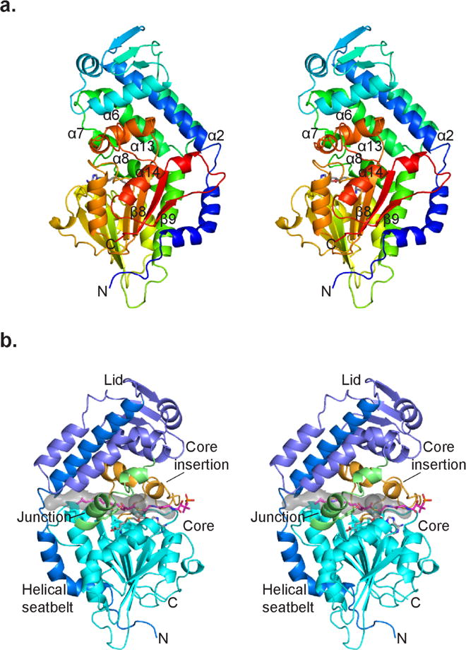
Figure 3.

CurJ C-MT structure. a) CurJ C-MT colored as a rainbow from blue (N-terminus) to red (C-terminus), shown in stereo. SAH is shown in sticks with atomic colors (C, gray; O, red; N, blue; S, yellow). b) CurJ C-MT colored by structure region. Helical seatbelt, blue; lid, dark blue; lid-to-core junction green; core, cyan; core insertion, orange; SAH, sticks with gray C. The transparent gray surface represents the substrate tunnel, which is lined with hydrophobic residues (Ile35, Phe157, Leu168, Val174, Ala307, Trp313, Val314, Phe318, Leu338). The CurJ C-MT substrate and phosphopantetheine (sticks with magenta C) were modeled into the active site. The views in A and B are from opposite sides of the C-MT.
