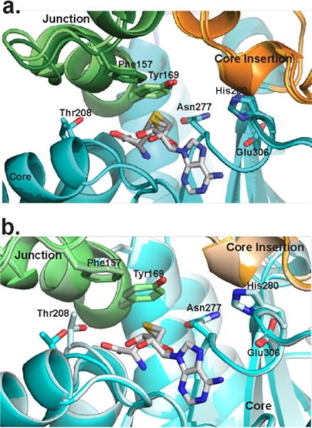
Figure 6.

Movement surrounding the CurJ C-MT active site. Structural regions are colored as in Fig. 3b. a) Closely related SeMet and wild type CurJ C-MT crystal forms (light and dark colors) are different in the lid-to-core junction and SAH homocysteine position. b) The position of Thr208 in MT motif I differs in the two C-MT chains in the SeMet crystal form.
