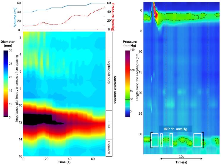
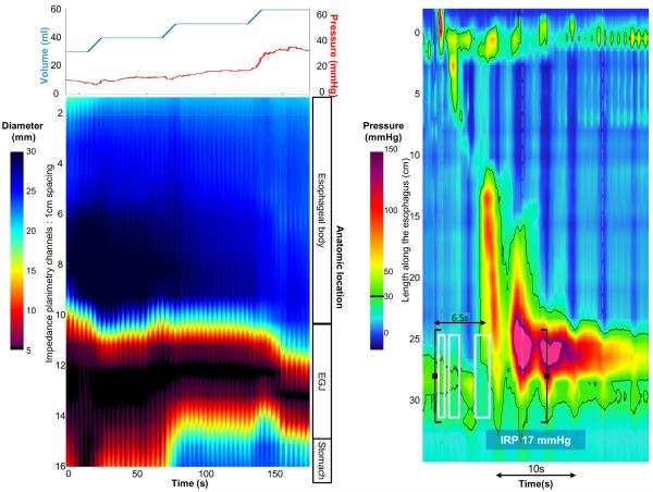
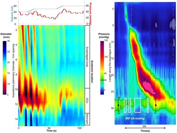
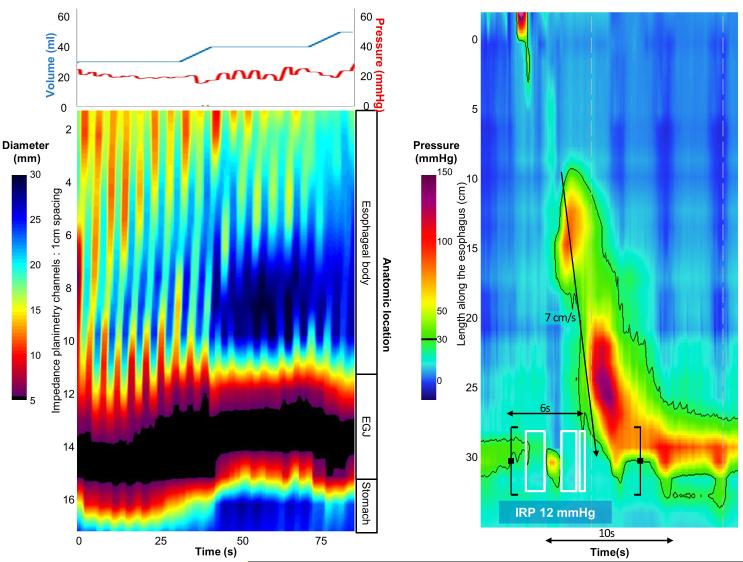
Figure 5. Case examples of FLIP topography.
Left panels represent portions of the FLIP study: distension volume (top, blue line), and intra-balloon pressure (top, red line), and FLIP topography (bottom). A swallow from the corresponding HRM is included in the right panels. A) A patient with type I achalasia, but borderline IRP (median 12 mmHg). FLIP topography demonstrated an abnormal EGJ-DI of 1.6 mm2/mmHg and absent contractility. B) A patient with EGJOO, and suspected evolving achalasia, on HRM. FLIP topography demonstrated abnormal EGJ-DI and absent contractility, supporting the diagnosis of achalasia. The patients was treated with a botulinum-toxin injection to the lower esophageal sphincter that resulted in improvement in dysphagia. C) A patient with EGJOO on HRM. FLIP topography was essentially normal with a normal EGJ-DI (4.9 mm2/mmHg) and RACs. An esophagram showed normal clearance of barium. Dysphagia had resolved without intervention at 6-month follow-up. D) A patient with normal motility on HRM (median IRP 13 mmHg), but with an abnormal EGJ-DI (0.46 mm2/mmHg) and RRCs on FLIP topography. Esophagram demonstrated persistent esophageal barium column height of 4.4-cm after 5 minutes and impaction of a 12.5 mm barium tablet at the EGJ. The patient underwent endoscopic ultrasound with diffusely thickened distal esophageal muscle layers supporting diagnosis of a primary esophageal motor disorder. A per-oral endoscopic myotomy was recommended


