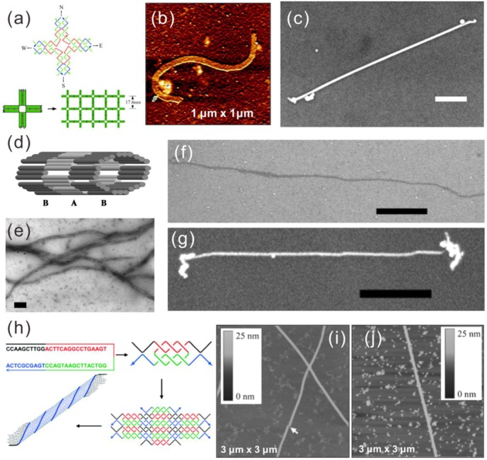
Figure 2.
Upper panel (a–c) [23]: (a) schematic of a 4 × 4 tile and nanoribbon assembly form from these tiles; (b) AFM image of a nanoribbon; (c) SEM image of a metallized silver nanoribbon, scale bar 500 nm; middle panel (d–g) [87]: (d) scheme of a nanotube made of TX tiles; (e,f) TEM and SEM image of nanotubes; (g) SEM image of a metallized silver nanotube; scale bars in (e–g) are 100 nm, 1 μm and 1 μm, respectively; lower panel (h–j) [24]: (h) assembly model of a nanotube from a single oligonucleotide with palindromic sequence, (i) AFM image of the nanotube; (j) metallized Pd nanotube. (a–c) are reproduced with permission from [23]. Copyright The American Association for the Advancement of Science, 2003; (d–g) are reproduced with permission from [87]. Copyright National Academy of Sciences, USA, 2004; (h–j) are reproduced with permission from [24]. Copyright John Wiley and Sons, 2006.

