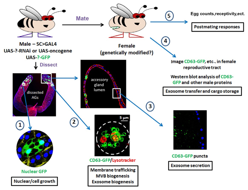
Fig. 3.
The secondary cell as a versatile in vivo model to study endolysosomal trafficking, exosome biogenesis, and functions. Diagram illustrates the different approaches that can be employed to study processes associated with exosome biogenesis and functions using the fly accessory glands, ranging from analysis of growth (1) and membrane trafficking/vesicle formation (2) in SCs, to exosome secretion into the gland lumen (3), to analysis of exosome transfer (4), and effects on postmating responses (5) in females. The living secondary cell shown in (2) is marked with CD63-GFP and stained with Lysotracker Red to reveal large acidic compartments, which, as in this case, are typically greater than 5 μm in diameter in older males. Note that the male and female can both be genetically modified in different ways to affect exosome-secreting and target cells, respectively. Exosomes and the compartments that produce them can also be imaged by more standard approaches, such as transmission electron microscopy (Corrigan et al., 2014).

