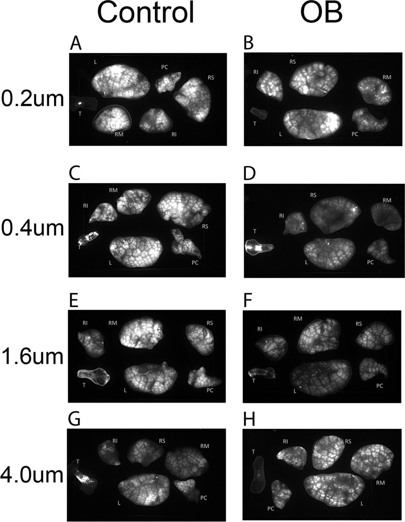
FIGURE 1.
(A) Control mouse lobes and trachea following exposure to the 0.2 µm aerosol, exposure time 200 ms. (B) OB mouse lobes and trachea following exposure to the 0.2 µm aerosol, exposure time 800 ms. (C) Control mouse lobes and trachea following exposure to the 0.4 µm aerosol, exposure time 200 ms. (D) OB mouse lobes and trachea following exposure to the 0.4 µm aerosol, exposure time 800 ms. (E) Control mouse lobes and trachea following exposure to the 1.0 µm aerosol, exposure time 100 ms. (F) OB mouse lobes and trachea following exposure to the 1.0 µm aerosol, exposure time 100 ms. (G) Control mouse lobes and trachea following exposure to the 4.0 µm aerosol, exposure time 200 ms. (H) OB mouse lobes and trachea following exposure to the 4.0 µm aerosol, exposure time 800 ms. Nomenclature for lobes is T: trachea, PC: postclaval, RM: right medial, RS: right superior, RI: right inferior, and L: left.

