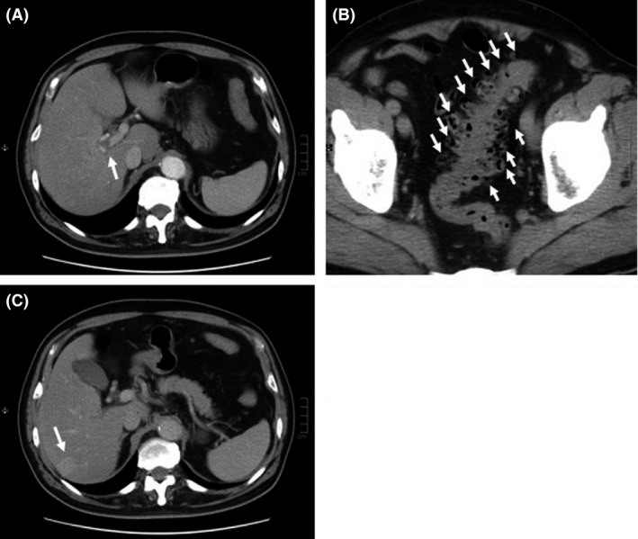
Figure 2.

Contrast‐enhanced computed tomography images of the abdomen on admission. (A) Intravascular thrombosis was observed in the right portal branch (white arrow). (B) Multiple diverticulae were also observed in the sigmoid colon (white arrows). (C) An approximately 2‐cm‐diameter lesion was observed in S7; this was slightly enhanced in the equilibrium phase.
