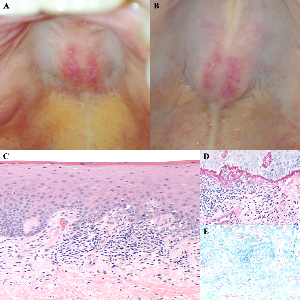
Figure 1.
Clinical photos of the ovoid palatal patch, with typical arcuate symmetric erythema on the hard palate intermixed with white macules (A,B). Biopsy of an ovoid palatal patch showed an interface dermatitis with dyskeratotic keratinocytes (C, 20× original magnification), a markedly thickened basement membrane (D, PASd, 40× original magnification), and increased dermal mucin (E, colloidal iron, 40× original magnification).

