Figure 1.
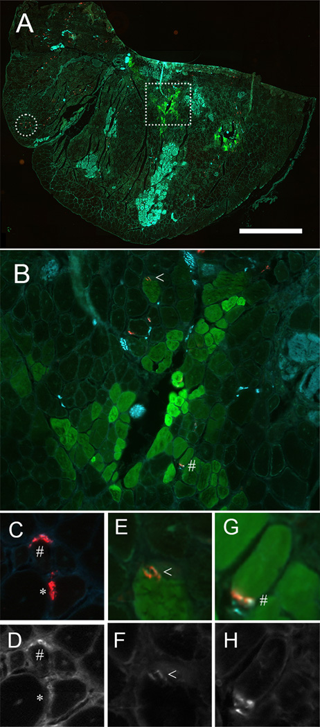
GFP+ muscle fibers are innervated. (A) Cryosection of the tibialis anterior muscle (TA) immunolabeled for GFP (green), postsynaptic acetylcholine receptors (AChR, red) and presynaptic vesicles and neurofilaments (cyan). (B) The region marked by the square in A is shown at higher magnification. (C–H) Paired color images of pre- and post-synaptic overlap along with greyscale images of presynaptic vesicles and neurofilament label. Neuromuscular junctions were categorized as denervated (*, no overlap between presynaptic label and AChR receptors above background), weak (<, diffuse overlap between presynaptic label and AChR receptors), or strong (#, bright punctate overlap between presynaptic label and AChR receptors). Fibers shown in C are from the area marked with a circle in A. Fibers shown in E & G are from area marked with a square in A and expanded in B. Scale bar = (A) 1000 µm, (B) 100 µm, and (C) 300 µm.

