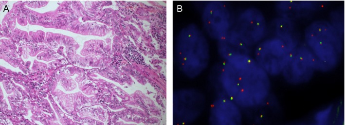
Figure 2.

Representative histopathological features (A) and break‐apart NRG1 FISH result (B) of the NRG1‐positive IMA case. (A) Goblet or columnar well differentiated tumoral cells with abundant intracytoplasmic mucin and small basally located nuclei (Hematoxylin‐Eosin‐Saffron, original magnification ×20) (B) Tumor nuclei hybridized with the ZytoLight® SPEC NRG1 dual color beak‐apart probe (ZytoVision). All tumor cell nuclei analyzed were positive, showing at least one isolated 3' (orange) signal. Original magnification ×630.
