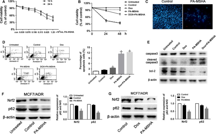
Figure 4.

PA‐MSHA inhibited MCF‐7/ADR cells proliferation through Nrf2/p62 in vitro. (A) MCF‐7/ADR cells were treated with various concentrations of PA‐MSHA for 12 h and 24 h, and cell viability was determined by the CCK8 assay. (B) The inhibitory effect of different drugs on MCF‐7/ADR cell proliferation. Cells were treated with PBS, doxorubicin (3 μg/mL), PA‐MSHA (0.848 × 10⁹cells/mL), and doxorubicin (3 μg/mL)+PA‐MSHA (0.848 × 10⁹cells/mL) for 48 h, and cell viability was determined by the CCK8 assay. (C) The effect of different drugs on MCF‐7/ADR cell apoptosis. Nucleus of MCF‐7/ADR cells were stained by Hoechst 33258. Cells were treated with PBS or PA‐MSHA (0.848 × 10⁹cells/mL) for 48 h. (D) MCF‐7/ADR cells were treated with PBS, doxorubicin (3 μg/mL), PA‐MSHA (0.848 × 10⁹cells/mL), and doxorubicin (3 μg/mL)+PA‐MSHA (0.848 × 10⁹cells/mL) for 48 h, and the apoptotic fraction of cells was detected by Annexin V staining/propidium iodide staining. (E) MCF7/ADR cells were treated with PBS, doxorubicin (3 μg/mL), PA‐MSHA (0.848 × 10⁹cells/mL), and doxorubicin (3 μg/mL)+PA‐MSHA (0.848 × 10⁹cells/mL) for 48 h, then the protein levels of caspase 3, cleaved‐caspase 3, and bcl‐2 were detected by western blot. (F) MCF7/ADR cells were treated with PBS, PA‐MSHA (0.848 × 10⁹cells/mL) for 8 h, then the protein levels of Nrf2 and p62 were detected by western blot. (G) MCF7/ADR cells were treated with PBS, doxorubicin (3 μg/mL), and PA‐MSHA (0.848 × 10⁹cells/mL) for 48 h, then the protein levels of Nrf2 and p62 were detected by western blot. PA‐MSHA, Pseudomonas aeruginosa mannose‐sensitive hemagglutinin; *P <0.05. PBS, phosphate‐buffered saline.
